Open to fellowship & radionuclide-therapy posts

Dr. Mohit Seth

MBBS · DNB Nuclear Medicine

Precision oncology through theranostics — imaging and treating cancer with the same molecule.

Delhi, India drmohitnm@gmail.com
Dr. Mohit Seth, Nuclear Medicine and Theranostics physician

Clinical experience at a glance

22,000+PET-CT studies reported / workload
168¹⁷⁷Lu-PSMA therapies (mCRPC)
92PRRT ¹⁷⁷Lu-DOTATATE (NET)
255I-131 post-therapy scans
2,300+SPECT-CT gamma studies
About

A Nuclear Medicine physician trained where theranostics is practised at scale — using molecular imaging to find cancer and targeted radionuclides to treat it.

Dr. Mohit Seth completed his DNB in Nuclear Medicine (2023–2026) at Rajiv Gandhi Cancer Institute & Research Centre (RGCIRC), Delhi — one of India's highest-volume theranostics centres. His practice spans the full precision-oncology pathway: Ga-68 PSMA, DOTANOC and FAPI PET/CT for staging, followed by ¹⁷⁷Lu-PSMA, ¹⁷⁷Lu-DOTATATE (PRRT) and I-131 radionuclide therapy, including hands-on management of inpatient therapy admissions.

He is first author on a prospective study of Ga-68 FAPI PET/CT in lung-cancer staging, presented orally at the SNMMI 2025 Annual Meeting in New Orleans, and is currently seeking an international fellowship in nuclear medicine and radionuclide therapy.

Expertise

Imaging and therapy, end to end

Diagnostic molecular imaging and the targeted radionuclide therapies that follow from it — the "see it, treat it" theranostics pairing, practised across a high-volume oncology service.

Radionuclide therapy

  • 168 · ¹⁷⁷Lu-PSMA (mCRPC)
  • 92 · ¹⁷⁷Lu-DOTATATE / PRRT (NET)
  • 255 · I-131 post-therapy scans (thyroid Ca)
  • I-131 mIBG · Y-90 radioembolisation
  • Inpatient therapy admission & isolation

Molecular imaging (PET/CT)

  • 22,000+ · PET-CT studies
  • 18F-FDG · Ga-68 PSMA · Ga-68 DOTANOC
  • Ga-68 FAPI · Ga-68 Trivehexin
  • F-18 DOPA
  • Staging, restaging & therapy response

Functional & SPECT studies

  • 2,300+ · gamma / SPECT-CT studies
  • Diuretic renography with GFR
  • Hepatobiliary & future-remnant-liver volumetry
  • Parathyroid · thyroid · bone · MUGA
  • Salivary-gland scintigraphy
Research & Publications

Original work in FAPI & PSMA imaging

First-author research and case work at the frontier of theranostics imaging — with a focus on FAPI PET/CT and incidental findings on PSMA studies.

First author Published · JNM

Incremental role of Ga-68 FAPI PET/CT for staging lung cancer evaluated for surgery: a prospective comparative study with 18F-FDG PET/CT

Seth M, Choudhury P, Darlong L, Gupta M, Gambhir A, Pathak A.

Journal of Nuclear Medicine 2025;66(suppl 1):252179 — presented at the SNMMI Annual Meeting, New Orleans. View abstract →

FAPI PET/CTSensitivity 84.6% · AUC 0.84
FDG PET/CTSensitivity 61.5% · AUC 0.72

Prospective series of 16 treatment-naive surgical candidates; specificity 75.6% for both. Ga-68 FAPI PET/CT was more sensitive than 18F-FDG PET/CT for nodal staging.
Full manuscript Under review

Extended manuscript of the Ga-68 FAPI lung-cancer staging study

The complete peer-reviewed paper expanding on the published abstract is currently under review.

Case reports Submitted

Multiple oncology case reports on PSMA and FDG PET/CT findings

Several first-author case reports documenting incidental malignancies and atypical presentations on molecular imaging — submitted and awaiting publication.

Presentations & Awards

Presented & recognised

Oral presentation — SNMMI 2025, New Orleans

Ga-68 FAPI PET/CT in lung-cancer staging (in person, Jun 2025).

Poster — SNMMI Mid-Winter & ACNM 2026, Orlando

Incidental non-prostatic malignancies on Ga-68 PSMA PET/CT.

Posters — AOCNMB 2025, Chennai

FAPI PET/CT in lung cancer; FDG PET/CT in follicular lymphoma.

Poster — ANMPICON 2025, Hyderabad

Incidental esophageal NEC unmasked on PSMA PET/CT.

Best Resident Award — RGCIRC, 2025

Department of Nuclear Medicine.

2nd position — ANMPICON 2025 Quiz

National nuclear medicine quiz.

Conferences & presenting

In the field

Active in the nuclear-medicine community — presenting research and representing the specialty at international meetings.

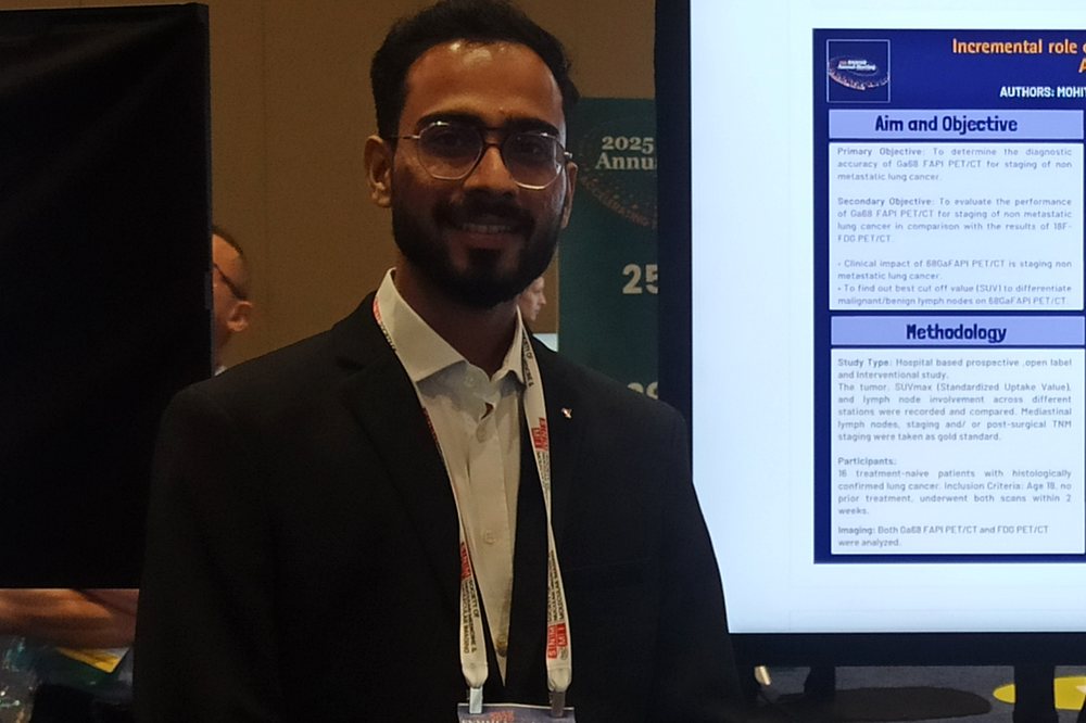
Dr. Mohit Seth presenting his Ga-68 FAPI PET/CT research poster at SNMMI 2025
Presenting Ga-68 FAPI research Oral presentation · SNMMI 2025, New Orleans
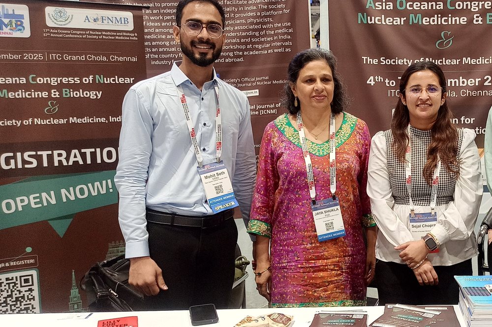
Dr. Mohit Seth with colleagues at the Society of Nuclear Medicine India community booth
Society of Nuclear Medicine India Community booth · SNMMI 2025
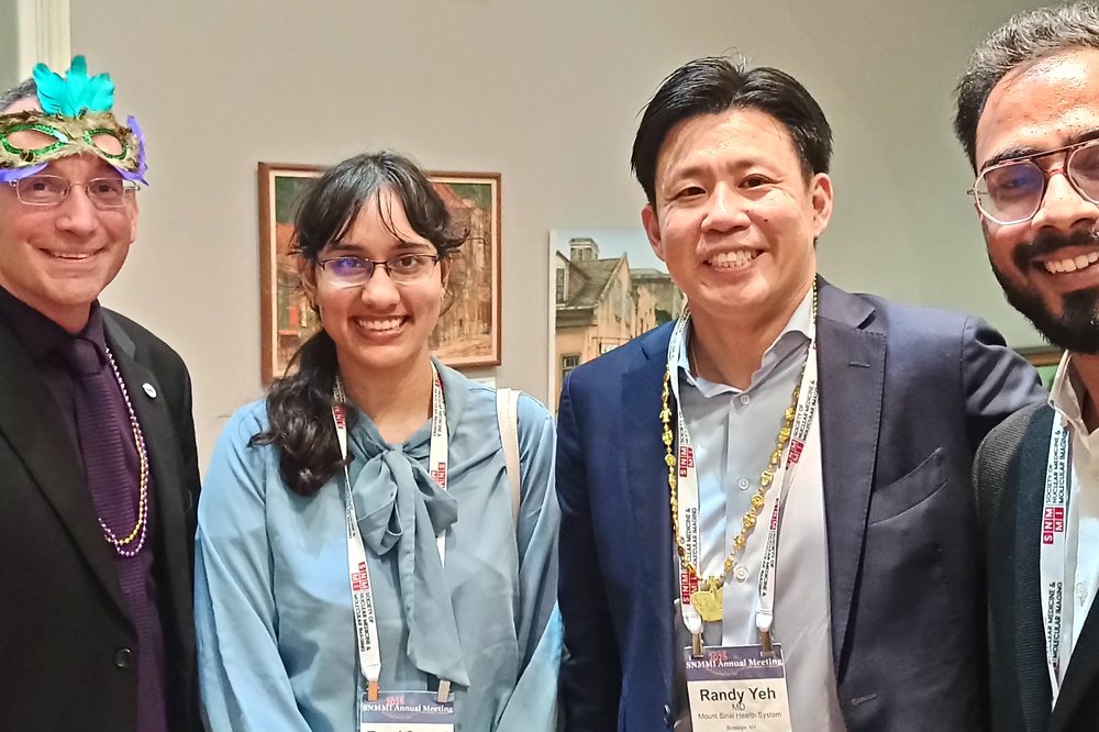
Dr. Mohit Seth with international colleagues at SNMMI 2025
With international colleagues SNMMI 2025, New Orleans
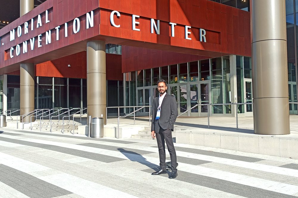
Dr. Mohit Seth at the Morial Convention Center for SNMMI 2025
SNMMI Annual Meeting Morial Convention Center · New Orleans
Education & Experience

Training & credentials

2026

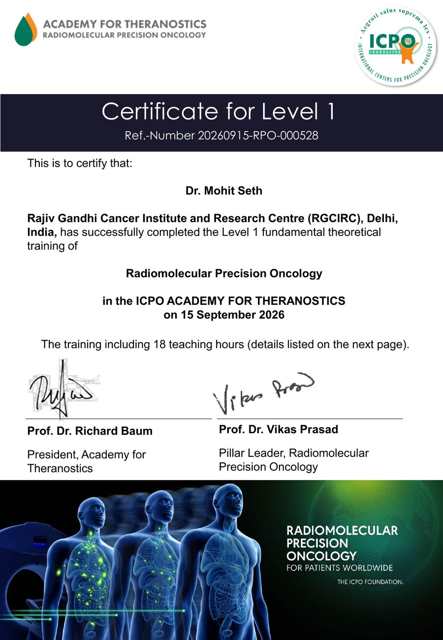
Certificate, Radiomolecular Precision Oncology (Level 1)

ICPO Academy for Theranostics — 18 hours of fundamental theoretical training in radiomolecular precision oncology. Signed by Prof. Dr. Richard Baum and Prof. Dr. Vikas Prasad.

Ref. 20260915-RPO-000528

ICPO Academy for Theranostics — Level 1 Radiomolecular Precision Oncology certificate for Dr. Mohit Seth View full certificate (PDF)
2023 — 2026

DNB, Nuclear Medicine

National Board of Examinations, New Delhi — trained at Rajiv Gandhi Cancer Institute & Research Centre (RGCIRC), Rohini, Delhi.

DNB Reg. No. 124-20139-231-249153

2023 — present

DNB Resident, Nuclear Medicine — RGCIRC

High-volume theranostics and molecular imaging service; inpatient radionuclide therapy management.

2023

Delhi Medical Council — Registration

Registered medical practitioner, Delhi Medical Council.

Reg. No. 111664 · registered 12 Dec 2023

2023

Medical Officer — Narayan Seva Sansthan, Udaipur

High-volume OPD/IPD care (≈140+ patients/day), May–Oct 2023.

MBBS

Bachelor of Medicine & Surgery

RNT Medical College, Udaipur.

Common questions

Nuclear medicine & theranostics, explained

Short answers to the questions patients, referring clinicians and colleagues ask most often about the imaging and therapies I work with.

Theranostics is the pairing of a diagnostic scan and a treatment that target the same molecule on a cancer cell. A radioactive tracer is first used to image where the target is present throughout the body; if the scan shows strong uptake, a therapeutic radionuclide attached to the same targeting molecule is then given to deliver radiation directly to those sites.

In practice this means the scan predicts who will benefit from the therapy — "see it, treat it" — which is why theranostics sits at the centre of precision oncology.

Ga-68 FAPI PET/CT images fibroblast activation protein, which is expressed by cancer-associated fibroblasts in the tumour's supporting tissue, whereas 18F-FDG PET/CT images glucose metabolism. Because FAPI targets the tumour stroma rather than glucose uptake, it can show lesions clearly in settings where FDG is limited — including tumours with low glucose avidity and areas of inflammation where FDG is less specific.

In our prospective study of lung cancer patients being evaluated for surgery, Ga-68 FAPI PET/CT was more sensitive than 18F-FDG PET/CT for nodal staging (84.6% vs 61.5%), with the same specificity. Read the published abstract →

¹⁷⁷Lu-PSMA is a radioligand therapy for metastatic castration-resistant prostate cancer, in which lutetium-177 is attached to a molecule that binds prostate-specific membrane antigen on prostate cancer cells. It delivers beta radiation directly to PSMA-expressing deposits throughout the body while largely sparing surrounding tissue.

Patients are selected using a Ga-68 PSMA PET/CT scan first — treatment is only appropriate where the scan confirms sufficient PSMA expression at sites of disease.

Peptide receptor radionuclide therapy, or PRRT, treats neuroendocrine tumours by attaching lutetium-177 to a somatostatin analogue that binds somatostatin receptors on tumour cells. It is given as a series of infusions, typically four cycles spaced roughly eight weeks apart.

As with PSMA therapy, eligibility is confirmed on imaging first — a Ga-68 DOTANOC or DOTATATE PET/CT scan must show adequate receptor expression.

Radioiodine therapy uses I-131, taken orally as a capsule or liquid, to treat differentiated thyroid cancer and some overactive thyroid conditions. Thyroid tissue naturally concentrates iodine, so the radioactive iodine is absorbed by remaining thyroid or thyroid-cancer cells and destroys them from within.

Because patients emit low levels of radiation afterwards, higher-dose treatment involves a short stay in a dedicated isolation room, followed by a whole-body scan a few days later to confirm where the iodine has localised.

Nuclear medicine images function — how tissue behaves biologically — while conventional radiology largely images structure. A CT or MRI shows the size and shape of a lesion; a PET or SPECT scan shows whether it is metabolically active or expressing a particular molecular target.

Nuclear medicine is also therapeutic: the same specialty that performs the scan administers targeted radionuclide treatment, which is what distinguishes it from purely diagnostic imaging.

Please note: this is general information about nuclear medicine procedures, not medical advice. Treatment decisions depend on individual circumstances and should always be made with your own treating team.

Let's talk

Open to fellowship and radionuclide-therapy positions. The fastest way to reach me is directly by email or phone.